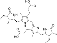
Stercobilin
Stercobilin is a tetrapyrrolic bile pigment and is one end-product of heme catabolism.[1][2] It is the chemical responsible for the brown color of human feces and was originally isolated from feces in 1932. Stercobilin (and related urobilin) can be used as a marker for biochemical identification of fecal pollution levels in rivers.[3]
 | |
| Names | |
|---|---|
| IUPAC name
3-[(2E)-2-[ [3-(2-Carboxyethyl)-5- [(4-ethyl-3-methyl-5-oxo-pyrrolidin-2-yl) methyl]-4-methyl-1H-pyrrol-2-yl]methylidene]-5- [(3-ethyl-4-methyl-5-oxo-pyrrolidin-2-yl) methyl]-4-methyl-pyrrol-3-yl]propanoic acid | |
| Identifiers | |
3D model (JSmol) |
|
| ChemSpider | |
| ECHA InfoCard | 100.047.155 |
| MeSH | Stercobilin |
PubChem CID |
|
| UNII | |
CompTox Dashboard (EPA) |
|
| |
| |
| Properties | |
| C33H46N4O6 | |
| Molar mass | 594.742 |
Except where otherwise noted, data are given for materials in their standard state (at 25 °C [77 °F], 100 kPa). | |
| Infobox references | |
Metabolism
Stercobilin results from breakdown of the heme moiety of hemoglobin found in erythrocytes. Macrophages break down senescent erythrocytes and break the heme down into biliverdin, which rapidly reduces to free bilirubin. Bilirubin binds tightly to plasma proteins (especially albumin) in the blood stream and is transported to the liver, where it is conjugated with one or two glucuronic acid residues into bilirubin diglucuronide, and secreted into the small intestine as bile. In the small intestine, some bilirubin glucuronide is converted back to bilirubin via bacterial enzymes in the terminal ileum. This bilirubin is further converted to colorless urobilinogen. Urobilinogen that remains in the colon can either be reduced to stercobilinogen and finally oxidized to stercobilin, or it can be directly reduced to stercobilin. Stercobilin is responsible for the brown color of human feces. Stercobilin is then excreted in the feces.[4]
Role in disease
Obstructive jaundice
In obstructive jaundice, no bilirubin reaches the small intestine, meaning that there is no formation of stercobilinogen. The lack of stercobilin and other bile pigments causes feces to become clay-colored.[4]
Brown pigment gallstones
An analysis of two infants suffering from cholelithiasis observed that a substantial amount of stercobilin was present in brown pigment gallstones. This study suggested that brown pigment gallstones could form spontaneously in infants suffering from bacterial infections of the biliary tract.[5]
Role in treatment of disease
A 1996 study by McPhee et al. suggested that stercobilin and other related pyrrolic pigments — including urobilin, biliverdin, and xanthobilirubic acid — has potential to function as a new class of HIV-1 protease inhibitors when delivered at low micromolar concentrations. These pigments were selected due to a similarity in shape to the successful HIV-1 protease inhibitor Merck L-700,417. Further research is suggested to study the pharmacological efficacy of these pigments.[6]
See also
References
- Boron W, Boulpaep E. Medical Physiology: A cellular and molecular approach, 2005. 984-986. Elsevier Saunders, United States. ISBN 1-4160-2328-3
- Kay IT, Weimer M, Watson CJ (1963). “The formation in vitro of stercobilin from bilirubin” Journal of Biological Chemistry. 238:1122-3. PMID 14031566
- Lam, Ching-Wan; Lai, Chi-Kong; Chan, Yan-Wo (1 February 1998). "Simultaneous Fluorescence Detection of Fecal Urobilins and Porphyrins by Reversed-Phase High-Performance Thin-Layer Chromatography". Clinical Chemistry. 44 (2): 345–346. doi:10.1093/clinchem/44.2.345. PMID 9474036.
- Seyfried H, Klicpera M, Leithner C, Penner E (1976). “Bilirubin metabolism”. Wiener Klinische Wochenschrift. 88:477-82. PMID 793184
- Treem, William R.; Malet, Peter F.; Gourley, Glenn R.; Hyams, Jeffrey S. (February 1989). "Bile and Stone Analysis in Two Infants With Brown Pigment Gallstones and Infected Bile". Gastroenterology. 96 (2): 519–523. doi:10.1016/s0016-5085(89)91579-5. PMID 2642880. Retrieved 3 June 2020.
- McPhee, Fiona; Caldera, Patricia S.; Bemis, Guy W.; McDonagh, Antony F.; Kuntz, Irwin D.; Craik, Charles S. (1 December 1996). "Bile pigments as HIV-1 protease inhibitors and their effects on HIV-1 viral maturation and infectivity in vitro". Biochemical Journal. 320 (2): 681–686. doi:10.1042/bj3200681. PMC 1217983. PMID 8973584.