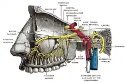
Tympanic nerve
The tympanic nerve (Jacobson’s nerve) is a branch of the glossopharyngeal nerve found near the ear.
| Tympanic nerve | |
|---|---|
 Plan of upper portions of glossopharyngeal, vagus, and accessory nerves. (Tympanic nerve visible in upper right) | |
 Tympanic nerve (labelled right side) | |
| Details | |
| To | tympanic plexus |
| Identifiers | |
| Latin | nervus tympanicus |
| TA98 | A14.2.01.138 |
| TA2 | 6323 |
| FMA | 53480 |
| Anatomical terms of neuroanatomy | |
Path
It arises from the inferior ganglion of the glossopharyngeal nerve, and ascends to the tympanic cavity through a small canal, the inferior tympanic canaliculus, on the under surface of the petrous portion of the temporal bone on the ridge which separates the carotid canal from the jugular fossa.
In the tympanic cavity it divides into branches which form the tympanic plexus and are contained in grooves upon the surface of the promontory.
The tympanic nerve contains both sensory and parasympathetic axons:
- Sensory axons innervate the middle ear including the internal surface of the tympanic membrane. Their cell bodies are found in the superior ganglion of the glossopharyngeal nerve.
- Parasympathetic axons continue as the lesser petrosal nerve and innervate the postganglionic parasympathetic neurons of the otic ganglion. These neurons then provide secretomotor innervation of the parotid gland via the auriculotemporal nerve
Clinical significance
This nerve may be involved by paraganglioma, in this location referred to as glomus jugulare or glomus tympanicum tumours.
Additional images
 Lesser petrosal nerve
References
This article incorporates text in the public domain from page 910 of the 20th edition of Gray's Anatomy (1918)
External links
- Tympanic and Lesser petrosal nerve diagram
- cranialnerves at The Anatomy Lesson by Wesley Norman (Georgetown University) (IX)