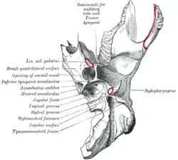
Carotid canal
The carotid canal is the passageway in the temporal bone through which the internal carotid artery enters the middle cranial fossa from the neck. The canal starts on the inferior surface of the temporal bone at the external opening of the carotid canal (also referred to as the carotid foramen). The canal ascends at first superiorly, and then, making a bend, runs anteromedially. The canal's internal opening is near the foramen lacerum, above which the internal carotid artery passes on its way anteriorly to the cavernous sinus.
| Carotid canal | |
|---|---|
 Left temporal bone. Inferior surface. ("Opening of carotid canal" labeled at center left.) | |
| Details | |
| Identifiers | |
| Latin | canalis caroticus |
| TA98 | A02.1.06.013 |
| TA2 | 651 |
| FMA | 55805 |
| Anatomical terms of bone | |
Contents
The carotid canal allows the internal carotid artery to pass into the cranium,[1] as well as the carotid plexus traveling on the artery.
The carotid plexus contains sympathetics to the head from the superior cervical ganglion They have several motor functions: raise the eyelid (superior tarsal muscle), dilate pupil (pupillary dilator muscle), innervate sweat glands of face and scalp and constricts blood vessels in the head.
Additional images
 Horizontal section of nasal and orbital cavities.
 Coronal section of right temporal bone.
 Carotid canal.
References
This article incorporates text in the public domain from page 143 of the 20th edition of Gray's Anatomy (1918)
- Maynard, Robert Lewis; Downes, Noel (2019-01-01), Maynard, Robert Lewis; Downes, Noel (eds.), "Chapter 7 - The Cardiovascular System", Anatomy and Histology of the Laboratory Rat in Toxicology and Biomedical Research, Academic Press, pp. 77–90, ISBN 978-0-12-811837-5, retrieved 2021-01-06
External links
- Atlas image: n3a8p1 at the University of Michigan Health System
- "Anatomy diagram: 34257.000-1". Roche Lexicon - illustrated navigator. Elsevier. Archived from the original on 2012-07-22.
- Photo at Winona.edu