Supratrochlear nerve
The supratrochlear nerve is a branch of the frontal nerve, itself a branch of the ophthalmic nerve. It provides sensory innervation to the skin of the forehead and upper eyelid.
| Supratrochlear nerve | |
|---|---|
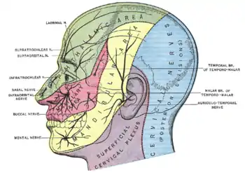
 Sensory areas of the head, showing the general distribution of the three divisions of the fifth nerve. (Supratrochlear nerve labeled at upper left.) | |
 Nerves of the orbit. Seen from above. (Supratrochlear nerve visible near top.) | |
| Details | |
| From | Frontal nerve |
| Identifiers | |
| Latin | nervus supratrochlearis |
| TA98 | A14.2.01.024 |
| TA2 | 6203 |
| FMA | 52642 |
| Anatomical terms of neuroanatomy | |
Structure
The supratrochlear nerve branches from the frontal nerve midway between the base and apex of the orbit. It travels anteriorly above the levator palpebrae superioris and exits the orbit through the supratrochlear notch in the superomedial margin of the orbit. It then ascends onto the forehead beneath the corrugator supercilii and frontalis muscles. It then divides into sensory branches.
Function
The supratrochlear nerve provides sensory innervation to the skin of the lateral forehead, upper eyelid and the conjunctiva.
Etymology
The supratrochlear nerve is named for its passage above the trochlea of the superior oblique muscle.
Additional images
 Supratrochlear nerve
 Extrinsic eye muscle. Nerves of orbita. Deep dissection.
 Extrinsic eye muscle. Nerves of orbita. Deep dissection.
 Extrinsic eye muscle. Nerves of orbita. Deep dissection.
 Extrinsic eye muscle. Nerves of orbita. Deep dissection.
 Extrinsic eye muscle. Nerves of orbita. Deep dissection.
References
This article incorporates text in the public domain from page 888 of the 20th edition of Gray's Anatomy (1918)
External links
- Anatomy figure: 29:02-01 at Human Anatomy Online, SUNY Downstate Medical Center
- MedEd at Loyola GrossAnatomy/h_n/cn/cn1/cnb1.htm
- lesson3 at The Anatomy Lesson by Wesley Norman (Georgetown University) (orbit2)
- cranialnerves at The Anatomy Lesson by Wesley Norman (Georgetown University) (V)
- http://www.dartmouth.edu/~humananatomy/figures/chapter_47/47-2.HTM