Septal nasal cartilage
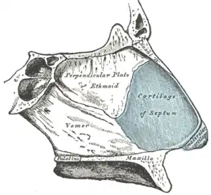
The septal nasal cartilage (cartilage of the septum or quadrangular cartilage) is composed of hyaline cartilage.[1] It is somewhat quadrilateral in form, thicker at its margins than at its center, and completes the separation between the nasal cavities in front.
| Septal nasal cartilage | |
|---|---|
 Bones and cartilages of septum of nose. Right side (cartilage of the septum visible as blue structure at right) | |
 Cartilages of the nose, seen from below (cartilage of septum visible in blue at bottom center) | |
| Details | |
| Identifiers | |
| Latin | Cartilago septi nasi |
| TA98 | A06.1.01.013 |
| TA2 | 946 |
| FMA | 59503 |
| Anatomical terminology | |
Its anterior margin, thickest above, is connected with the nasal bones, and is continuous with the anterior margins of the lateral cartilages; below, it is connected to the medial crura of the major alar cartilages by fibrous tissue.
Its posterior margin is connected with the perpendicular plate of the ethmoid; its inferior margin with the vomer and the palatine processes of the maxillae.
References
- Saladin, Kenneth S. Anatomy and Physiology (6th ed.). New York, NY: McGraw Hill, 2012.: McGraw Hill Higher Education. p. 856. ISBN 9780077472139.CS1 maint: location (link)
This article incorporates text in the public domain from page 992 of the 20th edition of Gray's Anatomy (1918)
External links
- Atlas image: rsa1p7 at the University of Michigan Health System - "Nasal septum, lateral view"
- Anatomy figure: 33:02-01 at Human Anatomy Online, SUNY Downstate Medical Center - "Diagram of skeleton of medial (septal) nasal wall."
- lesson9 at The Anatomy Lesson by Wesley Norman (Georgetown University) (nasalseptumbonescarti)