Romanowsky stain
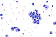
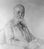
Romanowsky staining, also known as Romanowsky–Giemsa staining, is a prototypical staining technique that was the forerunner of several distinct but similar stains widely used in hematology (the study of blood) and cytopathology (the study of diseased cells). Romanowsky-type stains are used to differentiate cells for microscopic examination in pathological specimens, especially blood and bone marrow films,[1] and to detect parasites such as malaria within the blood.[2][3][4][5] Stains that are related to or derived from the Romanowsky-type stains include Giemsa, Jenner, Wright, Field, May–Grünwald and Leishman stains. The staining technique is named after the Russian physician Dmitri Leonidovich Romanowsky (1861–1921), who was one of the first to recognize its potential for use as a blood stain.[6]
.jpg.webp)
Mechanism
The value of Romanowsky staining lies in its ability to produce a wide range of hues, allowing cellular components to be easily differentiated. This phenomenon is referred to as the Romanowsky effect, or more generally as metachromasia.[7]
Romanowsky effect

In 1891 Romanowsky[8][9][10] developed a stain using a mixture of eosin (typically eosin Y) and aged solutions of methylene blue that formed hues unattributable to the staining components alone: distinctive shades of purple in the chromatin of the cell nucleus and within granules in the cytoplasm of some white blood cells. This became known as the Romanowsky or Romanowsky-Giemsa effect.[11][12][6][13][4] Eosin and pure methylene blue alone (or in combination) do not produce the Romanowsky effect,[13][4] and the active stains which produce the effect are now considered to be azure B and eosin.[14][3][13]
Polychromed methylene blue
Romanowsky-type stains can be made from either a combination of pure dyes, or from methylene blue that has been subject to oxidative demethylation, which results in the breakdown of methylene blue into multiple other stains, some of which are necessary to produce the Romanowsky effect.[15][4] Methylene blue that has that has undergone this oxidative process is known as "polychromed methylene blue".[15][4] Polychromed methylene blue may contain up to 11 dyes, including methylene blue, azure A, azure B, azure C, thionine, methylene violet Bernthesen, methyl thionoline and thionoline.[4] The exact composition of polychromed methylene blue depends on the method used, and even batches of the stain from the same manufacturer may vary in composition.[15]
Although azure B and eosin have been shown to be the required components to produce the Romanowsky effect,[14][3][13] these stains in their pure forms have not always been used in the formulation of the staining solutions.[4] The original sources of azure B (one of the oxidation products of methylene blue) were from polychromed methylene blue solutions, which were treated with oxidizing agents or allowed to naturally age in the case of Romanowsky.[3][13] Ernst Malachowsky in 1891 was the first to purposely polychrome methylene blue for use in a Romanowsky-type stain.[15][16]
Types
May-Grünwald-Giemsa
The May-Grünwald-Giemsa stain is a two step procedure that includes first staining with May-Grünwald stain, which does not produce the Romanowsky effect, followed by staining the Giemsa stain which does produce the Romanowsky effect.[17]
Wright's and Wright-Giemsa stains
Wright's stain can be used alone or in combination with the Giemsa stain, which is known as the Wright-Giemsa stain.[1] Wright's stain is named after James Homer Wright who in 1902[18] published a method using heat to produce polychromed methylene blue, which is combined with eosin Y.[19][20][21][1] The polychromed methylene blue is combined with eosin and allowed to precipitate, forming an eosinate which is redissolved in methanol.[4] The addition of Giemsa to Wright's stain increases the brightness of the "reddish-purple" color of the cytoplasmic granules.[1][21] The Wright's and Wright-Giemsa stains are two of the Romanowsky-type stains in common use in the United States and are mainly used for the staining of blood and bone marrow films.[21][1]
Leishman stain
In 1901 William Leishman[22] developed a stain that was similar to Louis Jenner's but with the replacement of pure methylene blue with polychromed methylene blue.[19][15][4] Leishman's stain is prepared from the eosinate of polychromed methylene blue and eosin Y using methanol as the solvent.[4]
Giemsa stain
Giemsa stain is composed of "Azure II" and eosin Y with methanol and glycerol as the solvent.[15] "Azure II" is thought to be a mixture of azure B (which Giemsa called "azure I") and methylene blue, although the exact composition of "azure I" is considered a trade secret.[4][15]Comparable formulations using known dyes have been published and are commercially available. Giemsa stain is considered to be the standard stain for detection and identification of the malaria parasite.[5]
Clinical importances
Blood and bone marrow pathology

Romanowsky-type stains are widely used in the examination of blood, in the form of blood films, and in the microscopic examination of bone marrow biopsies and aspirate smears.[1][23] Examination of both blood and bone marrow can be of importance in the diagnosis of a variety of blood diseases.[1][23] In the United States the Wright and Wright-Giemsa variants of the Romanowsky-type stains are widely used,[1] while in Europe Giemsa stain is commonly employed.[1]
Detection of malaria and other parasites
Of the Romanowsky-type stains, the Giemsa stain is especially important in the detection and identification of malaria parasites in blood samples.[5][15] Malaria antigen detection tests are an alternative to the staining and microscopic examination of blood films for the detection of malaria.[5]
Use in cytopathology
Romanowsky-type stains are also used for the staining of cytopathologic specimens such as those produced from fine-needle aspirates and cerebrospinal fluid from lumbar punctures.[24]
History
.jpg.webp)
Although debate exists as to who deserves credit for this general staining method, popular usage has attributed it to Dmitri Leonidovich Romanowsky.[14][16][19]
In the 1870s Paul Ehrlich used a mixture of acidic and basic dyes including acid fuchsin (acid dye) and methylene blue (basic dye) to examine blood films.[25][26][27][28][16] In 1888 Cheslav Ivanovich Chenzinsky used methylene blue, but substituted the acid fuchsin used by Ehrlich with eosin.[14][27][28] Chenzinsky's stain combination was able to stain the malaria parasite (a member of the genus Plasmodium).[28][19] Neither Ehrlich's or Chenzinsky's stains produced the Romanowsky effect as the methylene blue they used was not polychromed.[16]
Dmitri Romanowsky in 1890 published preliminary findings of his blood stain (a combination of aged methylene blue and eosin), including the results when applied to malaria infected blood.[6] This use of polychromed methylene blue differentiated Romanowsky's stain (and the subsequent formulations) from those of Ehrlich and Chenzinsky, which lacked the purple hue associated with the Romanowsky effect.[16] Romanowsky's 1890 publication did not include a description of how he modified his methylene blue solution,[6][16] but in his 1891 doctoral thesis he described methylene blue best as used after mold began forming on the surface.[6][16] Other than the use of an aged methylene blue solution, Romanowsky's stain was based on Chenzinsky's stain technique.[16] Romanowsky's use of his method to study the malaria patasite has been attributed to the continued interest in his staining method.[26]

Ernst Malachowsky has been credited with independently observing the same stain combination as Dmitri Romanowsky in 1891,[6][13] although he has also been credited with being the first to do so.[16][19] Malachowsky was the first to use a deliberately polychromed methylene blue solution,[15] which Malachowsky accomplished by the addition of borax to the staining mixture.[16] Malachowsky is reported to have demonstrated the stain on June 15, 1890, and in the same year to have published a paper "describing his public demonstration".[19] Both the Romanowsky and Malachowsky methods were able to stain the nucleus and cytoplasm of the malaria parasite, when until this point the stains used had only colored the cytoplasm.[19]
In 1899, Louis Leopold Jenner developed a more stable version of the methylene blue and eosin stain by collecting the precipitate that forms in water-based mixtures and redissolving it in methanol.[28][15][4] Romanowsky-type stains prepared from the collected precipitates are sometimes known as eosinates.[4] Besides increasing the stability of the stain, the use of methanol in Jenner's stain had the effect of fixing the blood samples,[4] although Jenner's version of the stain does not produce the Romanowsky effect.[28][19][15]
Richard May and Ludwig Grünwald in 1892 published a version of the stain (now known as the May–Grünwald stain) which is similar to the version proposed by Jenner in 1899, and likewise does not produce the Romanowsky effect.[28][19][15]
In 1901, both Karl Reuter and William Leishman[22] developed stains that combined Louis Jenner's use of alcohol as the solvent and Malachowsky's use of polychromed methylene blue.[19][15][4] Reuter's stain differed from Jenner's in using ethyl alcohol instead of methanol, and Leishman's differed from Jenner's by using eosin B instead of eosin Y.[19][4]
James Homer Wright in 1902 published[18] a method using heat to polychrome the methylene blue, which he combined with eosin Y. This technique is known as Wright's stain.[19][20]
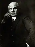
Gustav Giemsa's name has also become associated with the stain as he is credited with publishing a useful formulation and protocol in 1902.[13][6][26] Giemsa attempted to use combinations of pure dyes rather than polychromed methylene blue solutions which are highly variable in composition.[20][19][15] Giemsa sold the rights to produce his stain, but never fully published details on how he produced it,[19] although it is thought that he used a combination of azure B and methylene blue.[15] Giemsa published a number of modifications of his stains between 1902 and 1934. In 1904[29] he suggested adding glycerin to his stain, along with the methanol, to increase its stability.[23][19]
Giemsa stain powders produced in Germany were widely used in the United States until the interruption of the supply during World War I, which caused increased utilization of James Homer Wright's method for polychroming methylene blue.[19][1]
References
- Theil, Karl S. (2012). "Bone Marrow Processing and Normal Morphology". Laboratory Hematology Practice. pp. 279–299. doi:10.1002/9781444398595.ch22. ISBN 9781444398595.
- Bain, Barbara J.; Bates, Imelda; Laffan, Mike A. (11 August 2016). "Chapter 4: Preparation and staining methods for blood and bone marrow films". Dacie and Lewis Practical Haematology (12 ed.). Elsevier Health Sciences. ISBN 978-0-7020-6925-3.
- Horobin, RW (2011). "How Romanowsky stains work and why they remain valuable — including a proposed universal Romanowsky staining mechanism and a rational troubleshooting scheme". Biotechnic & Histochemistry. 86 (1): 36–51. doi:10.3109/10520295.2010.515491. ISSN 1052-0295.
- Marshall, P. N. (1978). "Romanowsky-type stains in haemotology". The Histochemical Journal. 10 (1): 1–29. doi:10.1007/BF01003411. PMID 74370.
- Li, Qigui; Weina, Peter J.; Miller, R. Scott (2012). "Malaria Analysis". Laboratory Hematology Practice. pp. 626–637. doi:10.1002/9781444398595.ch48. ISBN 9781444398595.
- Bezrukov, AV (2017). "Romanowsky staining, the Romanowsky effect and thoughts on the question of scientific priority". Biotechnic & Histochemistry. 92 (1): 29–35. doi:10.1080/10520295.2016.1250285. ISSN 1052-0295.
- Ribatti, Domenico (2018). "The Staining of Mast Cells: A Historical Overview". International Archives of Allergy and Immunology. 176 (1): 55–60. doi:10.1159/000487538. ISSN 1018-2438.
- Романовскiй Д.Л. (1890). "Къ вопросу о строенiи чужеядныхъ малярiи". Врачъ. 52: 1171–1173.
- Романовскiй Д.Л. Къ вопросу о паразитологіи и терапiи болотной лихорадки. Диссертацiя на степень доктора медицины. Спб. 1891 г., 118 с.
- Romanowsky D (1891). "Zur Frage der Parasitologie und Therapie der Malaria". St Petersburg Med Wochenschr. 16: 297–302, 307–315.
- Horobin RW, Walter KJ (1987). "Understanding Romanowsky staining. I: The Romanowsky-Giemsa effect in blood smears". Histochemistry. 86 (3): 331–336. doi:10.1007/bf00490267. PMID 2437082.
- Woronzoff-Dashkoff KK. (2002). "The wright-giemsa stain. Secrets revealed". Clin Lab Med. 22 (1): 15–23. doi:10.1016/S0272-2712(03)00065-9. PMID 11933573.
- Wittekind, D. H. (1983). "On the nature of Romanowsky-Giemasa staining and its significance for cytochemistry and histochemistry: an overall view". The Histochemical Journal. 15 (10): 1029–1047. doi:10.1007/BF01002498. ISSN 0018-2214.
- Cooksey, CJ (2017). "Quirks of dye nomenclature. 8. Methylene blue, azure and violet". Biotechnic & Histochemistry. 92 (5): 347–356. doi:10.1080/10520295.2017.1315775. ISSN 1052-0295.
- Lillie, Ralph Dougall (1977). H. J. Conn's Biological stains (9th ed.). Baltimore: Williams & Wilkins. pp. 692p.
- Lillie, R. D. (1978). "Romanowsky–Malachowski Stains the So-Called Romanowsky Stain: Malachowski's 1891 Use of Alkali Polychromed Methylene Blue for Malaria Plasmodia". Stain Technology. 53 (1): 23–28. doi:10.3109/10520297809111439.
- Piaton, E.; Fabre, M.; Goubin-Versini, I.; Bretz-Grenier, M-F.; Courtade-Saïdi, M.; Vincent, S.; Belleannée, G.; Thivolet, F.; Boutonnat, J.; Debaque, H.; Fleury-Feith, J.; Vielh, P.; Egelé, C.; Bellocq, J-P.; Michiels, J-F.; Cochand-Priollet, B. (October 2016). "Guidelines for May-Grünwald-Giemsa staining in haematology and non-gynaecological cytopathology: recommendations of the French Society of Clinical Cytology (SFCC) and of the French Association for Quality Assurance in Anatomic and Cytologic Pathology (AFA". Cytopathology. 27 (5): 359–368. doi:10.1111/cyt.12323.
- Wright JH (1902). "A Rapid Method for the Differential Staining of Blood Films and Malarial Parasites". J Med Res. 7 (1): 138–144. PMC 2105822. PMID 19971449.
- Krafts, KP; Hempelmann, E; Oleksyn, BJ (2011). "The color purple: from royalty to laboratory, with apologies to Malachowski". Biotechnic & Histochemistry. 86 (1): 7–35. doi:10.3109/10520295.2010.515490. ISSN 1052-0295.
- Gatenby, J. B.; Beams, H. W. (1950). The Microtomist's Vade-Mecum (11th ed.). Philadelphia: The Blackstone Company.
- Woronzoff-Dashkoff KK (2002). "The wright-giemsa stain. Secrets revealed". Clin Lab Med. 22 (1): 15–23. doi:10.1016/S0272-2712(03)00065-9. PMID 11933573.
- Leishman W (1901). "Note on a Simple and Rapid Method of Producing Romanowsky Staining in Malarial and other Blood Films". Br Med J. 2 (2125): 757–758. doi:10.1136/bmj.2.2125.757. PMC 2507168. PMID 20759810.
- Peterson, Powers; McNeill, Sheila; Gulati, Gene (2012). "Cellular Morphologic Analysis of Peripheral Blood". Laboratory Hematology Practice. pp. 10–25. doi:10.1002/9781444398595.ch2. ISBN 9781444398595.
- Krafts, KP; Pambuccian, SE (2011). "Romanowsky staining in cytopathology: history, advantages and limitations". Biotechnic & Histochemistry. 86 (2): 82–93. doi:10.3109/10520295.2010.515492. ISSN 1052-0295.
- Ehrlich P (1880). "Methodologische Beiträge zur Physiologie und Pathologie der verschiedenen Formen der Leukocyten" (PDF). Z Klin Med. 1: 553–560. Archived from the original (PDF) on 2011-07-19.
- Bracegirdle, Brain (1986). A history of microtechnique : the evolution of the microtome and the development of tissue preparation (2nd ed.). Lincolnwood, IL: Science Heritage Ltd. ISBN 978-0940095007.
- Krafts, Kristine; Hempelmann, Ernst; Oleksyn, Barbara J. (2011). "In search of the malarial parasite". Parasitology Research. 109 (3): 521–529. doi:10.1007/s00436-011-2475-4. ISSN 0932-0113.
- Woronzoff-Dashkoff, Kristine Pauline Krafts (1993). "The Ehrlich-Chenzinsky-Plehn-Malachowski-Romanowsky-Nocht-Jenner-May-Grünwald-Leishman-Reuter-Wright-Giemsa-Lillie-Roe-Wilcox Stain: The Mystery Unfolds". Clinics in Laboratory Medicine. 13 (4): 759–771. doi:10.1016/S0272-2712(18)30406-2. ISSN 0272-2712.
- Giemsa G (1904). "Eine Vereinfachung und Vervollkommnung meiner Methylenazur-Methylenblau-Eosin-Färbemethode zur Erzielung der Romanowsky-Nochtschen Chromatinfärbung". Centralbl F Bakt Etc. 37: 308–311.
