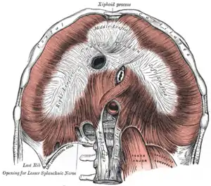
Lateral arcuate ligament
The lateral arcuate ligament (also lateral lumbocostal arch and external arcuate ligament) is a ligament under the diaphragm that arches across the upper part of the quadratus lumborum muscle. It is traversed by the subcostal nerve, artery and vein.
| Lateral arcuate ligament | |
|---|---|
 The diaphragm. Under surface. (Lat. arcuate ligament visible at bottom left.) | |
| Details | |
| Identifiers | |
| Latin | ligamentum arcuatum laterale |
| TA98 | A04.4.02.007 |
| TA2 | 2335, 2350 |
| FMA | 58283 |
| Anatomical terminology | |
Structure
The lateral arcuate ligament runs from the front of the transverse process of the first lumbar vertebra, and, laterally, to the tip and lower margin of the twelfth rib.[1] It forms an arch over the quadratus lumborum muscle.[1]
Variations
The lateral arcuate ligament is commonly described in anatomy textbooks as attaching at the first lumbar vertebra (L1).[2] However, other instances have been found in cadaver studies with attachments at either the second (L2) or third (L3) lumbar vertebra.[2]
In around 5% of people, inferolateral extensions of the lateral arcuate ligaments, such as thickened nodular areas, are found adjacent to the lateral diaphragmatic surface which can be visualized with computed tomography (CT) scans.[3]
History
The lateral arcuate ligaments were described by Galen, as early as AD 177.[4][5] This was found in his animal dissections performed as part of his Rome lectures, collected in De Anatomicus Administrationibus.[4][5]
References
This article incorporates text in the public domain from page 405 of the 20th edition of Gray's Anatomy (1918)
- Coakley, Fergus V.; Grant, Michael John; Behr, Spencer; Foster, Bryan R; Korngold, Elena K; Didier, Ryne A (2014-07-01). "Imaging of invasive thymoma in the costophrenic recess presenting as thickening of arcuate ligaments of the diaphragm". Clinical Imaging. 38 (4): 529–531. doi:10.1016/j.clinimag.2014.02.002. ISSN 0899-7071. PMC 4048795.
- Deviri E, Nathan H, Luchansky E (1988). "Medial and lateral arcuate ligaments of the diaphragm: attachment to the transverse process". Anat Anz. 166 (1–5): 63–7. PMID 3189849.
- Silverman PM, Cooper C, Zeman RK (1992). "Lateral arcuate ligaments of the diaphragm: anatomic variations at abdominal CT". Radiology. 185 (1): 105–8. doi:10.1148/radiology.185.1.1523290. PMID 1523290.
- Galen, Singer C (Trans.) "Galen on anatomical procedures: de Anatomicis administrationibus", Oxford University Press, 1956, p143.
- Derenne JP, Debru A, Grassino AE, Whitelaw WA (1995). "History of diaphragm physiology: the achievements of Galen". Eur. Respir. J. 8 (1): 154–60. doi:10.1183/09031936.95.08010154. PMID 7744182.
External links
- Anatomy figure: 40:04-10 at Human Anatomy Online, SUNY Downstate Medical Center - "The abdominal surface of the diaphragm."
- posteriorabdomen at The Anatomy Lesson by Wesley Norman (Georgetown University) (posteriorabdmus&nerves)