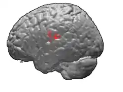
Brodmann area 43
Brodmann area 43, the subcentral area, is a structurally distinct area of the cerebral cortex defined on the basis of cytoarchitecture. Along with Brodmann Area 1, 2, and 3, Brodmann area 43 is a subdivision of the postcentral region of the brain,[1] suggesting a somatosensory ('feeling of the body') function. The histological structure of Area 43 was initially described by Korbinian Brodmann, but it was not labeled on his map of cortical areas.[2]
| Brodmann area 43 | |
|---|---|
 | |
 | |
| Details | |
| Identifiers | |
| Latin | Area subcentralis |
| NeuroNames | 1013 |
| NeuroLex ID | birnlex_1761 |
| FMA | 68640 |
| Anatomical terms of neuroanatomy | |
Location
In the human subcentral area 43, a sub area of the cytoarchitecture is defined in the postcentral region of the cerebral cortex. It occupies the postcentral gyrus, which is between the ventrolateral extreme of the central sulcus and the depth of the lateral sulcus, at the insula. Its rostral and caudal borders are approximated by the anterior subcentral sulcus and the posterior subcentral sulcus, respectively. Cytoarchitecturally, it is bounded rostrally, by the agranular frontal area 6, and caudally, for the most part, by the caudal postcentral area 2 and the supramarginal area 40.[1]
Function
Brodmann Area 43 responds to pressure on the eardrum and to oral intake (eating and drinking).[3] Because eating and drinking change pressure on the middle ear and eardrum, Brodmann Area 43 may be the primary somatosensory cortex of the eardrum.
Additionally, Brodmann Area 43 was found to be functionally active in a study differentiating the roles of the left-frontal and right-cerebellar regions during semantic analysis. Brodmann Area 43 showed a major increase in functional activation by fMRI, when study participants were asked to complete tasks which involved the selection of a verbal response from many possible responses, rather than a sustained search for a verbal response from few possible responses.[4]
In lower monkeys
Brodmann initially believed there to be no distinct Area 43 in his map of the lower monkey, the guenon. However, study of the myeloarchitecture of the region, by Theodor Mauss, determined that monkeys possess a structurally distinct area corresponding to the human subcentral area.[1] It was regarded as cytoarchitecturally homologous to area 30 of Mauss in 1908. However, research by Cécile and Oskar Vogt found no distinctive architectonic area of the corresponding location in the guenon.[5]
See also
References
- Garey LJ. Brodmann's Localisation in the Cerebral Cortex. New York : Springer, 2006 (ISBN 0-387-26917-7) (ISBN 978-0387-26917-7)
- Brodmann, Korobian. Vergleichende Lokalisationslehre der Grosshirnrinde: in ihren Prinzipien dargestellt auf Grund des Zellenbaues. Ja Barth, 1909.
- Job, Agnès (2011). "Cortical representation of tympanic membrane movements due to pressure variation: an fMRI study" (PDF). Hum. Brain Mapp. 32 (5): 744–9. doi:10.1002/hbm.21063. PMC 6870196. PMID 21484948.
- Gabrieli, John DE, Russell A. Poldrack, and John E. Desmond. "The role of left prefrontal cortex in language and memory." Proceedings of the national Academy of Sciences 95.3 (1998): 906-913.
- Vogt, Cécile, and Oskar Vogt. Allgemeine ergebnisse unserer hirnforschung. Vol. 25. JA Barth, 1919.
External links
| Wikimedia Commons has media related to Brodmann area 43. |
- For Neuroanatomy of the subcentral area 43 visit BrainInfo