Aryepiglottic fold
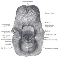
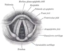
The aryepiglottic folds are triangular folds of mucous membrane enclosing ligamentous and muscular fibres. They are located at the entrance of the larynx, extending from the lateral borders of the epiglottis to the arytenoid cartilages, hence the name 'aryepiglottic'. They contain the aryepiglottic muscles and form the upper borders of the quadrangular membrane.
| Aryepiglottic fold | |
|---|---|
 The entrance to the larynx, viewed from behind (aryepiglottic fold labeled at center right) | |
 Laryngoscopic view of interior of larynx (aryepiglottic fold labeled at center right) | |
| Details | |
| Identifiers | |
| Latin | Plica aryepiglottica |
| TA98 | A06.2.09.003 |
| TA2 | 3193 |
| FMA | 55448 |
| Anatomical terminology | |
The folds are triangular in shape, being narrow in front, wide behind, and sloping obliquely downward and backward. They are bound, in front, by the epiglottis; behind, by the apices of the arytenoid cartilages, the corniculate cartilages, and the interarytenoid notch. Within the posterior part of each aryepiglottic fold exists a cuneiform cartilage which forms whitish prominence, the cuneiform tubercle. The aryepiglottic folds are shortened in laryngomalacia.
Phonatory mechanism
Under certain circumstances, the aryepiglottic folds take part in phonation, for instance in the singing technique of vocal growl, such as practiced by Louis Armstrong and other jazz singers. It has been proven that the approximation of the aryepiglottic folds during vocalization may establish sustained co-oscillations, at relatively low frequencies, producing the growl or growling effect.[1]
Gallery
 Aryepiglottic fold
 Aryepiglottic fold
 Aryepiglottic fold
 Aryepiglottic fold
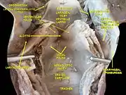
 Deep dissection of larynx, pharynx and tongue seen from behind
References
- Sakakibara, Ken-Ichi; Fuks, Leonardo; Imagawa, Hiroshi; Tayama, Niro (2004). "Growl Voice in Ethnic and Pop Styles" (PDF). Proceedings of the International Symposium on Musical Acoustics. Retrieved 19 June 2013.
- This article incorporates text in the public domain from page 1079 of the 20th edition of Gray's Anatomy (1918)
External links
- Anatomy photo:31:17-0104 at the SUNY Downstate Medical Center
- Atlas image: rsa3p10 at the University of Michigan Health System
- lesson11 at The Anatomy Lesson by Wesley Norman (Georgetown University) (larynxsagsect)