ACAT1
Acetyl-CoA acetyltransferase, mitochondrial, also known as acetoacetyl-CoA thiolase, is an enzyme that in humans is encoded by the ACAT1 (Acetyl-Coenzyme A acetyltransferase 1) gene.[5]
Acetyl-Coenzyme A acetyltransferase 1 is an acetyl-CoA C-acetyltransferase enzyme.
Structure
The gene spans approx. 27 kb and contains twelve exons interrupted by eleven introns. The region flanking the 5’ end of the gene lacks a TATA box, but contains many GC’s and also has two CAAT boxes. The gene also may have a binding site for the transcription factor Sp1, and has sequences resembling the binding sites of several other transcription factors. Additionally, there is a 101-bp DNA fragment immediately upstream from the cap site that has promoter activity.[6]
The human ACAT1 gene produces a chimeric mRNA through trans-splicing, a process in which separate transcripts from chromosomes 1 and 7 are spliced together. The chimeric mRNA transcript uses two sections to initiate translation: AUG(1397-1399) and GGC(1274-1276). Initiation of the first codon (AUG) results in the translation of a 50-kDa ACAT1, and initiation of the other (GGC) produces another enzymatically active 56-kDa isoform respectively; the 56kDa isoform is naturally present in human cells, including human monocyte-derived macrophages.[7]
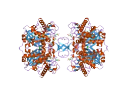
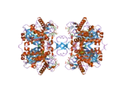
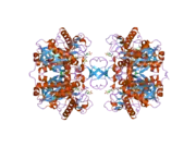
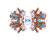
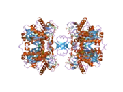
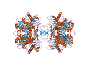
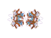
The resulting transcript encodes ACAT1, which is a 45.1 kDa protein composed of 427 amino acids.[8][9] It is also a homotetrameric protein that has nine transmembrane domains (TMDs). One active residue is a Histidine at the 460th position, which is in the 7th TMD. ACAT1 has seven free Cysteine residues, but they do not affect catalytic activity. There are two functional sections of this protein, TMD7 and TMD8; one side is involved in substrate binding and catalysis, while the other is involved in subunit interactions and binding.[10]
Function
This gene encodes a mitochondrially localized enzyme that catalyzes the reversible formation of acetoacetyl-CoA from two molecules of acetyl-CoA.[5] The ACAT1 enzyme has a few unique properties. First, it is activated by potassium ions binding near the CoA binding site and the catalytic site. This binding causes a structural change in the active site loop. Additionally, this enzyme is able to use 2-methyl-branched acetoacetyl-CoA as a substrate, making it a unique thiolase.[11] ACAT1 is regulated at both transcriptional and translational levels. ACAT1 enzyme activity is enhanced ACAT1’s expression is promoted transcriptionally by leptin,[12] angiotensin II,[13] and insulin in human monocytes/macrophages.[14] Insulin-mediated regulation also involves ERK, p38MAPK, and JNK signaling pathways.[15]
Clinical significance
Ketothiolase deficiency
Mutations of the ACAT1 gene are associated with a deficiency in the encoded protein mitochondrial acetoacetyl-CoA thiolase; this is also known as ketothiolase deficiency. Many mutations have been identified in specific populations, and large scale studies have been performed to determine the allelic and genotypic frequency for the defective gene.[16] As mitochondrial acetoacetyl-CoA thiolase is involved in beta-oxidation, a deficiency in this enzyme is marked by an increased amount of cholesterol compounds. Additionally, the isoleucine amino acid pathway is affected, such that proper metabolism of it is halted. This deficiency belongs to a more general class of disorders known as organic acidemias, in which the dysfunction of a specific step of amino acid catabolism results in the excretion of non-amino acids in the urine. This deficiency specifically presents as ketosis, acidosis, as well as hypoglycemia, but there are other clinical manifestations as well. The characteristics of organic acidemia disorders are vomiting, poor feeding, neurologic symptoms such as seizures and abnormal tone, and lethargy progressing to coma, which are all manifestations of toxic encephalopathy. The clinical outcome of infants with these disorders is largely determined by the time of diagnosis, with the potential outcome greatly improving if the disease is diagnosed in the first ten days of life. Ketothiolase deficiency is diagnosed by performing GC-MS and quantitative amino acid analysis in the urine; the diagnostic markers are 2-methyl-3-hydroxybutyric acid, 2-methylacetoacetic acid, and tiglylglycine. The disease is managed by trying to restore biochemical and physiologic homeostasis; common therapies include restricting diet to avoid the precursor amino acids and use of compounds to either dispose of toxic metabolites or increase enzyme activity. This disease is inherited in an autosomal recessive manner, meaning that carriers of the gene do not show symptoms of the disease.[17]
References
- GRCh38: Ensembl release 89: ENSG00000075239 - Ensembl, May 2017
- GRCm38: Ensembl release 89: ENSMUSG00000032047 - Ensembl, May 2017
- "Human PubMed Reference:". National Center for Biotechnology Information, U.S. National Library of Medicine.
- "Mouse PubMed Reference:". National Center for Biotechnology Information, U.S. National Library of Medicine.
- "Entrez Gene: acetyl-Coenzyme A acetyltransferase 1".
- Kano, M; Fukao, T; Yamaguchi, S; Orii, T; Osumi, T; Hashimoto, T (30 December 1991). "Structure and expression of the human mitochondrial acetoacetyl-CoA thiolase-encoding gene". Gene. 109 (2): 285–90. doi:10.1016/0378-1119(91)90623-j. PMID 1684944.
- Chen, J; Zhao, XN; Yang, L; Hu, GJ; Lu, M; Xiong, Y; Yang, XY; Chang, CC; Song, BL; Chang, TY; Li, BL (September 2008). "RNA secondary structures located in the interchromosomal region of human ACAT1 chimeric mRNA are required to produce the 56-kDa isoform". Cell Research. 18 (9): 921–36. doi:10.1038/cr.2008.66. PMC 3086790. PMID 18542101.
- Zong NC, Li H, Li H, Lam MP, Jimenez RC, Kim CS, Deng N, Kim AK, Choi JH, Zelaya I, Liem D, Meyer D, Odeberg J, Fang C, Lu HJ, Xu T, Weiss J, Duan H, Uhlen M, Yates JR, Apweiler R, Ge J, Hermjakob H, Ping P (Oct 2013). "Integration of cardiac proteome biology and medicine by a specialized knowledgebase". Circulation Research. 113 (9): 1043–53. doi:10.1161/CIRCRESAHA.113.301151. PMC 4076475. PMID 23965338.
- "Protein Information: P24752". Cardiac Organellar Protein Atlas Knowledgebase (COPaKB). Archived from the original on 14 August 2016. Retrieved 23 July 2016.
- Guo, ZY; Chang, CC; Chang, TY (4 September 2007). "Functionality of the seventh and eighth transmembrane domains of acyl-coenzyme A:cholesterol acyltransferase 1". Biochemistry. 46 (35): 10063–71. doi:10.1021/bi7011367. PMID 17691824.
- Haapalainen, AM; Meriläinen, G; Pirilä, PL; Kondo, N; Fukao, T; Wierenga, RK (10 April 2007). "Crystallographic and kinetic studies of human mitochondrial acetoacetyl-CoA thiolase: the importance of potassium and chloride ions for its structure and function". Biochemistry. 46 (14): 4305–21. doi:10.1021/bi6026192. PMID 17371050.
- Hongo, S; Watanabe, T; Arita, S; Kanome, T; Kageyama, H; Shioda, S; Miyazaki, A (August 2009). "Leptin modulates ACAT1 expression and cholesterol efflux from human macrophages". American Journal of Physiology. Endocrinology and Metabolism. 297 (2): E474–82. doi:10.1152/ajpendo.90369.2008. PMID 19625677.
- Kanome, T; Watanabe, T; Nishio, K; Takahashi, K; Hongo, S; Miyazaki, A (September 2008). "Angiotensin II upregulates acyl-CoA:cholesterol acyltransferase-1 via the angiotensin II Type 1 receptor in human monocyte-macrophages". Hypertension Research. 31 (9): 1801–10. doi:10.1291/hypres.31.1801. PMID 18971559.
- Ge, J; Zhai, W; Cheng, B; He, P; Qi, B; Lu, H; Zeng, Y; Chen, X (September 2013). "Insulin induces human acyl-coenzyme A: cholesterol acyltransferase1 gene expression via MAP kinases and CCAAT/enhancer-binding protein α.". Journal of Cellular Biochemistry. 114 (9): 2188–98. doi:10.1002/jcb.24568. PMID 23564383. S2CID 22816300.
- Xin, C; Yan-Fu, W; Ping, H; Jing, G; Jing-Jing, W; Chun-Li, M; Wei, L; Bei, C (May 2009). "Study of the insulin signaling pathways in the regulation of ACAT1 expression in cultured macrophages". Cell Biology International. 33 (5): 602–6. doi:10.1016/j.cellbi.2009.02.011. PMID 19269342. S2CID 13605913.
- Francis, T; Wartofsky, L (1 September 1992). "Common thyroid disorders in the elderly". Postgraduate Medicine. 92 (3): 225–30, 233–6. doi:10.1080/00325481.1992.11701452. PMID 1518756.
- Seashore, MR; Pagon, RA; Adam, MP; Ardinger, HH; Bird, TD; Dolan, CR; Fong, CT; Smith, RJH; Stephens, K (1993). "The Organic Acidemias: An Overview". Gene Reviews (R) Seattle (WA): University of Washington, Seattle; 1993-2015. Cite journal requires
|journal=(help) - Saraon, P; Trudel, D; Kron, K; Dmitromanolakis, A; Trachtenberg, J; Bapat, B; van der Kwast, T; Jarvi, KA; Diamandis, EP (April 2014). "Evaluation and prognostic significance of ACAT1 as a marker of prostate cancer progression". The Prostate. 74 (4): 372–80. doi:10.1002/pros.22758. PMID 24311408. S2CID 2169465.
- Saraon, P; Cretu, D; Musrap, N; Karagiannis, GS; Batruch, I; Drabovich, AP; van der Kwast, T; Mizokami, A; Morrissey, C; Jarvi, K; Diamandis, EP (June 2013). "Quantitative proteomics reveals that enzymes of the ketogenic pathway are associated with prostate cancer progression". Molecular & Cellular Proteomics. 12 (6): 1589–601. doi:10.1074/mcp.m112.023887. PMC 3675816. PMID 23443136.
External links
- Human ACAT1 genome location and ACAT1 gene details page in the UCSC Genome Browser.
Further reading
- Locke JA, Wasan KM, Nelson CC, et al. (2008). "Androgen-mediated cholesterol metabolism in LNCaP and PC-3 cell lines is regulated through two different isoforms of acyl-coenzyme A:Cholesterol Acyltransferase (ACAT)". Prostate. 68 (1): 20–33. doi:10.1002/pros.20674. PMID 18000807. S2CID 40860952.
- Fukao T, Boneh A, Aoki Y, Kondo N (2008). "A novel single-base substitution (c.1124A>G) that activates a 5-base upstream cryptic splice donor site within exon 11 in the human mitochondrial acetoacetyl-CoA thiolase gene". Mol. Genet. Metab. 94 (4): 417–21. doi:10.1016/j.ymgme.2008.04.014. PMID 18511318.
- Reynolds CA, Hong MG, Eriksson UK, et al. (2010). "Analysis of lipid pathway genes indicates association of sequence variation near SREBF1/TOM1L2/ATPAF2 with dementia risk". Hum. Mol. Genet. 19 (10): 2068–78. doi:10.1093/hmg/ddq079. PMC 2860895. PMID 20167577.
- Haapalainen AM, Meriläinen G, Pirilä PL, et al. (2007). "Crystallographic and kinetic studies of human mitochondrial acetoacetyl-CoA thiolase: the importance of potassium and chloride ions for its structure and function". Biochemistry. 46 (14): 4305–21. doi:10.1021/bi6026192. PMID 17371050.
- Chen J, Zhao XN, Yang L, et al. (2008). "RNA secondary structures located in the interchromosomal region of human ACAT1 chimeric mRNA are required to produce the 56-kDa isoform". Cell Res. 18 (9): 921–36. doi:10.1038/cr.2008.66. PMC 3086790. PMID 18542101.
- Raman J, Fritz TA, Gerken TA, et al. (2008). "The catalytic and lectin domains of UDP-GalNAc:polypeptide alpha-N-Acetylgalactosaminyltransferase function in concert to direct glycosylation site selection". J. Biol. Chem. 283 (34): 22942–51. doi:10.1074/jbc.M803387200. PMC 2517002. PMID 18562306.
- Xin C, Yan-Fu W, Ping H, et al. (2009). "Study of the insulin signaling pathways in the regulation of ACAT1 expression in cultured macrophages". Cell Biol. Int. 33 (5): 602–6. doi:10.1016/j.cellbi.2009.02.011. PMID 19269342. S2CID 13605913.
- Li Q, Bai H, Fan P (2008). "[Analysis of acyl-coenzyme A: cholesterol acyltransferase 1 polymorphism in patients with endogenous hypertriglyceridemia in Chinese population]". Zhonghua Yi Xue Yi Chuan Xue Za Zhi. 25 (2): 206–10. PMID 18393248.
- Guo ZY, Chang CC, Chang TY (2007). "Functionality of the seventh and eighth transmembrane domains of acyl-coenzyme A:cholesterol acyltransferase 1". Biochemistry. 46 (35): 10063–71. doi:10.1021/bi7011367. PMID 17691824.
- Fukao T, Yamaguchi S, Orii T, Hashimoto T (1995). "Molecular basis of beta-ketothiolase deficiency: mutations and polymorphisms in the human mitochondrial acetoacetyl-coenzyme A thiolase gene". Hum. Mutat. 5 (2): 113–20. doi:10.1002/humu.1380050203. PMID 7749408. S2CID 36280301.
- Barbe L, Lundberg E, Oksvold P, et al. (2008). "Toward a confocal subcellular atlas of the human proteome". Mol. Cell. Proteomics. 7 (3): 499–508. doi:10.1074/mcp.M700325-MCP200. PMID 18029348.
- Fukao T, Nguyen HT, Nguyen NT, et al. (2010). "A common mutation, R208X, identified in Vietnamese patients with mitochondrial acetoacetyl-CoA thiolase (T2) deficiency". Mol. Genet. Metab. 100 (1): 37–41. doi:10.1016/j.ymgme.2010.01.007. PMID 20156697.
- Hongo S, Watanabe T, Arita S, et al. (2009). "Leptin modulates ACAT1 expression and cholesterol efflux from human macrophages". Am. J. Physiol. Endocrinol. Metab. 297 (2): E474–82. doi:10.1152/ajpendo.90369.2008. PMID 19625677.
- Antalis CJ, Arnold T, Lee B, et al. (2009). "Docosahexaenoic acid is a substrate for ACAT1 and inhibits cholesteryl ester formation from oleic acid in MCF-10A cells". Prostaglandins Leukot. Essent. Fatty Acids. 80 (2–3): 165–71. doi:10.1016/j.plefa.2009.01.001. PMID 19217763.
- Bzoma B; Debska-Slizieñ A; Dudziak M; et al. (2008). "[Genetic predisposition to systemic complications of arterial hypertension in maintenance haemodialysis patients]". Pol. Merkur. Lekarski. 25 (147): 209–16. PMID 19112833.
- Kanome T, Watanabe T, Nishio K, et al. (2008). "Angiotensin II upregulates acyl-CoA:cholesterol acyltransferase-1 via the angiotensin II Type 1 receptor in human monocyte-macrophages". Hypertens. Res. 31 (9): 1801–10. doi:10.1291/hypres.31.1801. PMID 18971559.
- Ruaño G, Bernene J, Windemuth A, et al. (2009). "Physiogenomic comparison of edema and BMI in patients receiving rosiglitazone or pioglitazone". Clin. Chim. Acta. 400 (1–2): 48–55. doi:10.1016/j.cca.2008.10.009. PMID 18996102.
- Thümmler S, Dupont D, Acquaviva C, et al. (2010). "Different clinical presentation in siblings with mitochondrial acetoacetyl-CoA thiolase deficiency and identification of two novel mutations". Tohoku J. Exp. Med. 220 (1): 27–31. doi:10.1620/tjem.220.27. PMID 20046049.
- An S, Jang YS, Park JS, et al. (2008). "Inhibition of acyl-coenzyme A:cholesterol acyltransferase stimulates cholesterol efflux from macrophages and stimulates farnesoid X receptor in hepatocytes". Exp. Mol. Med. 40 (4): 407–17. doi:10.3858/emm.2008.40.4.407. PMC 2679275. PMID 18779653.
- Ewing RM, Chu P, Elisma F, et al. (2007). "Large-scale mapping of human protein-protein interactions by mass spectrometry". Mol. Syst. Biol. 3 (1): 89. doi:10.1038/msb4100134. PMC 1847948. PMID 17353931.
This article incorporates text from the United States National Library of Medicine, which is in the public domain.
