Pit (botany)
Pits are relatively thinner portions of the cell wall that adjacent cells can communicate or exchange fluid through. Pits are characteristic of cell walls with secondary layers. Generally each pit has a complementary pit opposite of it in the neighboring cell. These complementary pits are called "pit pairs".[1]

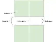
Pits are composed of three parts: the pit chamber, the pit aperture, and the pit membrane. The pit chamber is the hollow area where the secondary layers of the cell wall are absent. The pit aperture is the opening at either end of the pit chamber. The pit membrane is the primary cell wall and middle lamella, or the membrane between adjacent cell walls, at the middle of the pit chamber.[2]
The primary cell wall at the pit membrane may also have depressions similar to the pit depressions of the secondary layers. These depressions are primary pit-fields, or primary pits. In the primary pit, the primordial pit provides an interruption in the primary cell wall that the plasmodesmata can cross. The primordial pit is the only aperture in the otherwise continuous primary cell wall.[3]
Pit pairs are a characteristic feature of xylem, as sap flows through the pits of xylem cells.[4]
Types of pits

Though pits are usually simple and complementary, a few more pit variations can be formed:
- Simple pits: A pit pair in which the diameter of the pit chamber and the diameter of the pit aperture are equal.
- Bordered pits: A pit pair in which the pit chamber is over-arched by the cell wall, creating a larger pit chamber and smaller pit aperture.
- Half bordered pits: A pit pair in which a bordered pit has a complementary simple pit. Such a pit pair is called half bordered pit pair.
- Blind pits: A pit pair in which a simple pit has no complementary pit.
- Compound pits: A pit pair in which one cell wall has a large pit and the adjacent cell wall has numerous, small pits.
Plasmodesmata
Plasmodesmata are thin sections of the endoplasmic reticulum that traverse pits and connect adjacent cells. These sections provide an avenue of transport through the pits and facilitate communication.[5] Plasmodesmata are not restricted to pits however, as plasmodesmata often cross a cell wall of constant width and occasionally the cell wall is even wider in areas where plasmodesmata traverse it.[3]
Torus and margo

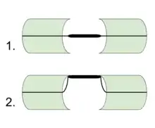
The torus and margo are characteristic features of bordered pit-pairs in gymnosperms, such as Coniferales, Ginkgo, and Gnetales. In other vascular plants, the torus is rare. The pit membrane is separated into two parts: a thick impermeable torus at the center of the pit membrane, and the permeable margo surrounding it. The torus regulates the functions of the bordered pit, and the margo is a cell wall-derived porous membrane that supports the torus. The margo is composed of bundles of microfibrils that radiate from the torus.[3]
The margo is flexible and can move towards either side of the pit while under stress. This allows the thick, impermeable torus to block the pit aperture. When the torus is displaced so that it blocks the pit aperture, the pit is said to be aspirated.
References
- Jeremy Burgess (1985). Introduction to Plant Cell Development. CUP Archive. p. 91. ISBN 9780521316118.
- Ray F. Evert (2006). Esau's Plant Anatomy: Meristems, Cells, and Tissues of the Plant Body: Their Structure, Function, and Development (third, illustrated ed.). John Wiley & Sons. p. 76. ISBN 9780470047378.
- Katherine Easu (1977). Anatomy of Seed Plants. Plant Anatomy (2nd ed.). John Wiley & Sons. p. 51. ISBN 0-471-24520-8.
- B. A. Meylan, Brian Geoffrey Butterfield (1972). Three-dimensional Structure of Wood: A Scanning Electron Microscope Study; Volume 2 of Syracuse wood science series (illustrated ed.). Syracuse University Press. p. 12. ISBN 9780815650300.
- Charles B. Beck (2010). An Introduction to Plant Structure and Development: Plant Anatomy for the Twenty-First Century (second, revised ed.). Cambridge University Press. p. 31. ISBN 9781139486361.
Further reading
| Wikimedia Commons has media related to Wood anatomy. |
- Andreas Bresinsky, Christian Körner, Joachim W. Kadereit, Gunther Neuhaus, Uwe Sonnewald: Strasburger – Lehrbuch der Botanik. Begründet von E. Strasburger. Spektrum Akademischer Verlag, Heidelberg 2008 (36. Aufl.) ISBN 978-3-8274-1455-7
- Dietger Grosser: Die Hölzer Mitteleuropas – Ein mikrophotographischer Holzatlas, Springer Verlag, 1977. ISBN 3-540-08096-1
- Rudi Wagenführ: Holzatlas, 6. neu bearb. und erw. Aufl., Fachbuchverlag Leipzig im Carl Hanser Verlag, München, 2007. ISBN 978-3-446-40649-0