ATP5D
ATP synthase subunit delta, mitochondrial, also known as ATP synthase F1 subunit delta or F-ATPase delta subunit is an enzyme that in humans is encoded by the ATP5F1D (formerly ATP5D) gene.[5][6][7] This gene encodes a subunit of mitochondrial ATP synthase. Mitochondrial ATP synthase catalyzes ATP synthesis, utilizing an electrochemical gradient of protons across the inner membrane during oxidative phosphorylation.[8]
Structure
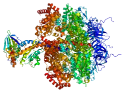
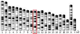
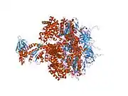
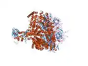
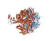
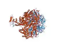
The ATP5F1D gene is located on the p arm of chromosome 19 at position 13.3 and it spans 3,075 base pairs.[8] The ATP5F1D gene produces a 17.5 kDa protein composed of 168 amino acids.[9][10] The coded protein is a subunit of the mitochondrial ATP synthase (Complex V), which is composed of two linked multi-subunit complexes: the soluble catalytic core, F1, and the membrane-spanning component, Fo, comprising the proton channel. The catalytic portion of mitochondrial ATP synthase consists of 5 different subunits (alpha, beta, gamma, delta, and epsilon) assembled with a stoichiometry of 3 alpha, 3 beta, and a single representative of the other 3. The proton channel consists of three main subunits (a, b, c). This gene encodes the delta subunit of the catalytic core. Alternatively spliced transcript variants encoding the same isoform have been identified.[8] The structure of the protein has been known to resemble a 'lollipop' structure due to the attachment of the F1 catalytic unit to the mitochondrial inner membrane by the F0 unit.[11]
Function
This gene encodes a subunit of the mitochondrial ATP synthase (Complex V) of the mitochondrial respiratory chain, which is necessary for the catalysis of ATP synthesis. Utilizing an electrochemical gradient of protons produced by electron transport complexes of the respiratory chain, the synthase converts ADP into ATP across the inner membrane during oxidative phosphorylation.[8] F-type ATPases consist of two structural domains, F1 and F0, that contribute to the catalysis. The F1 domain contains an extramembranous catalytic core and the F0 domain contains the membrane proton channel linked by a central and a peripheral stalk. During catalysis, ATP turnover in the catalytic domain of F1 is coupled by a rotary mechanism of the central stalk subunits to proton transport. The encoded protein is a part of the complex F1 domain and of the central stalk which is part of the complex rotary element. Rotation of the central stalk against the surrounding alpha3beta3 subunits leads to the hydrolysis of ATP in three separate catalytic sites on the beta subunits.[5][6]
Clinical significance
Mutations of ATP5F1D have been associated with childhood mitochondrial disorders with phenotypes such as episodic decompensations, lactic acidosis, and hyperammonemia accompanied by ketoacidosis or hypoglycemia. Biallelic mutations of c.245C>T and c.317T>G in ATP5F1D were shown to cause a metabolic disorder with such phenotypes due to mitochondrial dysfunction in two unrelated individuals.[12] Mutations of ATP5F1D with decreased expression of the protein has also been found to result in synaptic dysfunction of the mitochondria that could play an essential role in amyotrophic lateral sclerosis (ALS) pathogenesis.[13]
Interactions
Among the two components, CF1 - the catalytic core - and CF0 - the membrane proton channel of the F-type ATPase, ATP5F1D is associated with the catalytic core. The catalytic core is composed of five different subunits including alpha, beta, gamma, delta, and epsilon subunits. The protein has additional interactions with ATP5I, ATP5O, PUS1, NDUFB5, GTPBP6, ATP5L, ATP5J and others.[14][5][6]
References
- GRCh38: Ensembl release 89: ENSG00000099624 - Ensembl, May 2017
- GRCm38: Ensembl release 89: ENSMUSG00000003072 - Ensembl, May 2017
- "Human PubMed Reference:". National Center for Biotechnology Information, U.S. National Library of Medicine.
- "Mouse PubMed Reference:". National Center for Biotechnology Information, U.S. National Library of Medicine.
-
"ATP5F1D - ATP synthase subunit delta, mitochondrial precursor - Homo sapiens (Human) - ATP5F1D gene & protein". Retrieved 2018-08-07.
 This article incorporates text available under the CC BY 4.0 license.
- "UniProt: the universal protein knowledgebase". Nucleic Acids Research. 45 (D1): D158–D169. January 2017. doi:10.1093/nar/gkw1099. PMC 5210571. PMID 27899622.
- Jordan EM, Breen GA (February 1992). "Molecular cloning of an import precursor of the delta-subunit of the human mitochondrial ATP synthase complex". Biochimica et Biophysica Acta (BBA) - Gene Structure and Expression. 1130 (1): 123–6. doi:10.1016/0167-4781(92)90477-h. PMID 1531933.
- "Entrez Gene: ATP5D ATP synthase, H+ transporting, mitochondrial F1 complex, delta subunit".
 This article incorporates text from this source, which is in the public domain.
 - Zong NC, Li H, Li H, Lam MP, Jimenez RC, Kim CS, et al. (October 2013). "Integration of cardiac proteome biology and medicine by a specialized knowledgebase". Circulation Research. 113 (9): 1043–53. doi:10.1161/CIRCRESAHA.113.301151. PMC 4076475. PMID 23965338.
- "ATP synthase subunit delta, mitochondrial". Cardiac Organellar Protein Atlas Knowledgebase (COPaKB).
- Walker JE (May 1995). "Determination of the structures of respiratory enzyme complexes from mammalian mitochondria". Biochimica et Biophysica Acta (BBA) - Molecular Basis of Disease. 1271 (1): 221–7. doi:10.1016/0925-4439(95)00031-x. PMID 7599212.
- Oláhová M, Yoon WH, Thompson K, Jangam S, Fernandez L, Davidson JM, et al. (March 2018). "Biallelic Mutations in ATP5F1D, which Encodes a Subunit of ATP Synthase, Cause a Metabolic Disorder". American Journal of Human Genetics. 102 (3): 494–504. doi:10.1016/j.ajhg.2018.01.020. PMC 6117612. PMID 29478781.
- Engelen-Lee J, Blokhuis AM, Spliet WG, Pasterkamp RJ, Aronica E, Demmers JA, et al. (May 2017). "Proteomic profiling of the spinal cord in ALS: decreased ATP5D levels suggest synaptic dysfunction in ALS pathogenesis". Amyotrophic Lateral Sclerosis & Frontotemporal Degeneration. 18 (3–4): 210–220. doi:10.1080/21678421.2016.1245757. PMID 27899032.
- Mick DU, Dennerlein S, Wiese H, Reinhold R, Pacheu-Grau D, Lorenzi I, Sasarman F, Weraarpachai W, Shoubridge EA, Warscheid B, Rehling P (2012). "MITRAC links mitochondrial protein translocation to respiratory-chain assembly and translational regulation". Cell. 151 (7): 1528–41. doi:10.1016/j.cell.2012.11.053. PMID 23260140.
Further reading
- Yoshida M, Muneyuki E, Hisabori T (September 2001). "ATP synthase--a marvellous rotary engine of the cell". Nature Reviews. Molecular Cell Biology. 2 (9): 669–77. doi:10.1038/35089509. PMID 11533724. S2CID 3926411.
- Hochstrasser DF, Frutiger S, Paquet N, Bairoch A, Ravier F, Pasquali C, Sanchez JC, Tissot JD, Bjellqvist B, Vargas R (December 1992). "Human liver protein map: a reference database established by microsequencing and gel comparison". Electrophoresis. 13 (12): 992–1001. doi:10.1002/elps.11501301201. PMID 1286669. S2CID 23518983.
- Yasuda R, Noji H, Kinosita K, Yoshida M (June 1998). "F1-ATPase is a highly efficient molecular motor that rotates with discrete 120 degree steps". Cell. 93 (7): 1117–24. doi:10.1016/S0092-8674(00)81456-7. PMID 9657145. S2CID 14106130.
- Wang H, Oster G (November 1998). "Energy transduction in the F1 motor of ATP synthase". Nature. 396 (6708): 279–82. Bibcode:1998Natur.396..279W. doi:10.1038/24409. PMID 9834036. S2CID 4424498.
- Cross RL (January 2004). "Molecular motors: turning the ATP motor". Nature. 427 (6973): 407–8. Bibcode:2004Natur.427..407C. doi:10.1038/427407b. PMID 14749816. S2CID 52819856.
- Itoh H, Takahashi A, Adachi K, Noji H, Yasuda R, Yoshida M, Kinosita K (January 2004). "Mechanically driven ATP synthesis by F1-ATPase". Nature. 427 (6973): 465–8. Bibcode:2004Natur.427..465I. doi:10.1038/nature02212. PMID 14749837. S2CID 4428646.
External links
- Human ATP5D genome location and ATP5D gene details page in the UCSC Genome Browser.
This article incorporates text from the United States National Library of Medicine, which is in the public domain.
